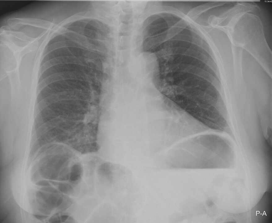
A 60-year-old woman presents with a one-week history of abdominal pain, nausea and reduced frequency of bowel motions. Her observations are as follows: HR 97, BP 113/76, RR 19, SaO2 98% on air. On examination she has right upper quadrant tenderness. Her chest X-ray is shown below:

1. Which sign is present on her chest X-ray?
Show Answer
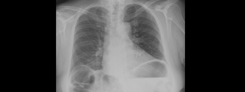
Chilaiditi sign is present on this X-ray, which is the presence of visible gas below the right diaphragm. Rugal folds are often seen within the gas showing that it lies within the bowel and is not free. Chilaiditi sign is caused by the interposition of a loop of large intestine between the diaphragm and the liver.
Image sourced from Wikipedia
Courtesy of Steven Fruitsmaak CC BY-SA 3.0
2. What is the most likely diagnosis?
Show Answer
This patient has a diagnosis of Chiliaditi syndrome. Most cases where Chilaiditi sign is present patients are completely asymptomatic, but in cases where symptoms are present, it is referred to as Chilaiditi syndrome.
The symptoms of Chilaiditi syndrome are extremely variable with some patients experiencing only mild intermittent abdominal pain, whilst others present with severe pain that can precipitate hospitalisation. Other symptoms that can occur include constipation, vomiting, dysphagia, breathlessness. Rarely volvulus can occur, causing intestinal obstruction.
3. Which conditions can predispose to this diagnosis?
Show Answer
The exact cause of Chilaiditi syndrome is unknown but it occurs with greater frequency in individuals with:
- Dolichocolon (long and mobile colon)
- Chronic obstructive pulmonary disease
- Liver cirrhosis
- Ascites
4. How is this condition usually managed?
Show Answer
Asymptomatic patients without symptoms require no specific treatment. Those patients that develop abdominal pain are generally managed conservatively with analgesia and fluid resuscitation.
Occasionally patients with intractable symptoms or those that develop bowel ischaemia will require surgical intervention. Procedures that are used under these circumstances include transverse colectomy, right hemicolectomy and hepatopexy (anchoring of displaced liver to the abdominal wall).


Great case
Loved the details n presentations?
The case is beautiful and the explanation is thorough and very clear.
Unusual case. Thank you!