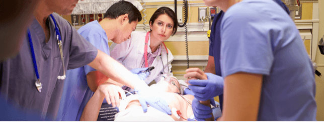
The initial assessment and management of the seriously injured trauma patient is both challenging and anxiety-inducing for many clinicians. It is an undertaking that requires a cool head, systematic approach, speed, and good clinical judgement.
The Advanced Trauma Life Support (ATLS) program was introduced in the 1980s to address the need for higher-quality trauma care, particularly in the “first hour” after injury, following an incident in which an orthopaedic surgeon crashed his plane in a rural setting. The surgeon sustained serious injuries, three of his children sustained critical injuries, and one child sustained minor injuries. Sadly, his wife died instantly in the crash. He felt that the care that he and his family received was sub-standard, stating at the time:
“When I can provide better care in the field with limited resources than what my children and I received at the primary care facility, there is something wrong with this system, and the system has to be changed.”
This led to the development of the ATLS program that is so widely attended around the world today.
The Trauma Team
The ATLS program is designed so that a single doctor can safely look after a patient with multiple serious injuries by performing tasks in a stepwise, systematic approach. In reality, however, most hospitals have a fully equipped and prepared ‘Trauma Team’, each member with an allocated role, allowing each of these stepwise tasks to be performed more quickly, and often simultaneously.
A typical trauma team will be composed of the following key members:
- Dedicated team leader
- Anaesthetist or airway management expert
- Anaesthetic assistant
- General surgeon
- Orthopaedic surgeon
- Emergency physician(s)
- Nursing staff (generally a minimum of 2)
- Emergency radiographer
- Scribe
The ‘ABCDE’ Approach
The ATLS approach to the management of the seriously injured trauma patient starts with a rapid primary survey, using an ‘ABCDE’ approach. This allows life-threatening injuries to be identified in a prioritized sequence and treated accordingly.
The sequence is as follows:
- Airway maintenance with cervical spine protection
- Breathing and ventilation
- Circulation with haemorrhage control
- Disability: Neurologic status
- Exposure/Environmental control
Airway maintenance with cervical spine protection
The first priority when evaluating a trauma patient is to assess the airway to ensure that it is patent. The patient should be inspected externally, looking for signs of facial injury, foreign bodies, blood, and vomit. The patient should then be asked to state their name, and attention should be paid to noisy breathing and audible signs of imminent airway obstruction. If a patient can communicate clearly verbally, then is unlikely that the airway is in immediate jeopardy. Suctioning and Magill’s forceps should be used to assist in clearing the airway if necessary.
If airway manoeuvers are required, then the chin-lift or jaw thrust manoeuvers are recommended, as these also protect the cervical spine. Airway adjuncts may also be required, with nasopharyngeal airways recommended in conscious patients. In unconscious patients, oropharyngeal airways can be used as a temporary measure in patients with no gag reflex. A definitive airway should be established in patients that are unable to maintain their airway integrity. The mental status of the patient should be quickly assessed, and a Glasgow Coma Scale (GCS) score of 8 or less usually requires the placement of a definitive airway. In patients with severe facial and/or neck injuries a surgical airway, e.g. cricothyroidotomy or emergency tracheostomy, may be necessary.
The ATLS program strongly advocates cervical spine protection throughout the airway management phase and entire primary survey. The theory behind this is that the use of cervical collars and spinal immobilisation adjuncts prevents secondary spinal cord injury during the assessment and transport of trauma patients. This practice has come under some scrutiny in recent times, however, with some clinicians suggesting that this practice is not evidence-based and that there is no need to immobilise the cervical spine of an awake, alert patient without neurological deficits or complaints. A nice review of this subject can be found here on the R.E.B.E.L EM site.
While this is an interesting topic to follow, it should be stressed that the current ATLS guidelines still state that cervical spine protection is required and this is the current gold standard in trauma care.
Breathing and ventilation
The next step in the evaluation of the trauma patient is the assessment of their breathing and ventilation. All trauma patients should receive high-flow oxygen via a non-rebreather mask during the primary survey.
The patient’s chest should be fully exposed, and the chest and neck inspected carefully looking at jugular venous distension, the position of the trachea, signs of external trauma, and asymmetrical movement of the chest. The entire chest wall should be palpated looking for signs of chest injury that could compromise ventilation, including surgical emphysema and crepitus. Chest percussion can also identify abnormalities but can be difficult in a noisy resuscitation area. The chest should then be carefully auscultated, listening for air entry bilaterally, gauging adequacy, and assessing for added sounds.
Any injuries discovered that severely impair ventilation should be addressed and managed at this stage of the primary survey. These potentially life-threatening injuries can be easily remembered using the ‘ATOM-FC’ mnemonic:
- Airway disruption
- Tension pneumothorax
- Open pneumothorax
- Massive haemothorax
- Flail chest
- Cardiac tamponade
Circulation with haemorrhage control
The next step in the evaluation of the trauma patient is the assessment of the circulation. Blood loss is the main preventable cause of death after injury, and the identification and control of haemorrhage is therefore of vital importance. The patient should be carefully inspected for any sources of major external bleeding, and the level of consciousness, skin colour and pulse should be assessed.
The major areas of internal bleeding are the chest, abdomen, retroperitoneum, pelvis, and long bones. The source of bleeding can usually be established through a combination of a thorough clinical examination, and trauma imaging. The imaging traditionally used in the primary survey has been chest and pelvic radiography, but increasingly this is being augmented with the use of focused assessment sonography in trauma (FAST scanning). The immediate management of life-threatening internal haemorrhage may include chest decompression, the use of pelvic binders, the application of splints, and surgical intervention.
Disability: neurological status
Following the circulatory assessment, a rapid neurological evaluation should be performed. The patient’s conscious level should be formally assessed using the GCS, and the pupils should be inspected for size, symmetry and reaction to light.
The patient should also be examined for signs of spinal cord injury, and if present the level of the spinal cord injury established.
It should be noted that oxygenation, ventilation, drugs and alcohol, and hypoglycaemia can also affect the level of consciousness, and this should be borne in mind during this disability assessment.
Exposure and environmental control
The final step in the primary survey is to fully expose the patient. All clothing should be removed or cut off to facilitate a thorough examination and assessment.
Once the patient has been exposed the patient should be covered with a warm blanket or external warming device to prevent hypothermia. Any intravenous fluids or blood products that are administered should also be warmed before infusion.
The resuscitation area should be a warm environment, and it is important to remember that the patient’s body temperature is far more important than the comfort of the healthcare providers that make up the trauma team.
Once the primary survey is completed, resuscitative efforts underway, and the patient’s vital functions have been normalised, the next stage in the initial evaluation of the trauma patient is to perform the secondary survey.
Next: The Initial Trauma Assessment Part 2 – The Secondary Survey
Header image used on licence from Shutterstock
Thank you to the joint editorial team of www.mrcemexamprep.net for this article.


After reading the article i am of the opinion that every medical graduate should know the managemement of severely injured patients keeping in view the advanced trauma life support.
need of every doctor to learn ATLS.