The optic nerve (CN II) is the second of the cranial nerves. It transmits visual sensory information from the retina to the brain. It passes from the optic disc to the optic chiasm, and then continues as the optic tract to the lateral geniculate nucleus, and then to the primary visual cortex of the occipital lobe.
In order to gain a full understanding of the optic nerve, it is essential to understand the anatomy of the retina and the entire visual pathway.
The retina
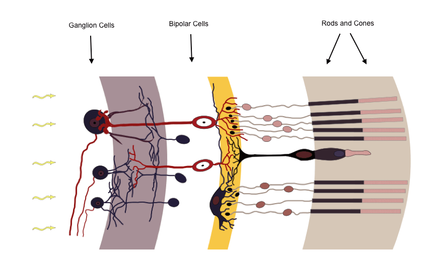
The retina is the light-sensitive inner layer of the eye. It is a layered structure of neurons that are interconnected by synapses. The only neurons that are light sensitive are the photoreceptor cells, the rods and cones (shown on the far right of the image below).
The rods function mainly in dim light and are responsible for black-and-white vision. The cones function best in bright light and are responsible for colour vision. When light reaches the rods and cones, a chemical change occurs that results in a signal being transmitted to the horizontal layer (the yellow layer in the image below) and then onto the bipolar cells (red cells on the image below).
The bipolar cells are situated between the photoreceptor cells and the ganglion cells. They have a central body from which two sets of processes arise that synapse either with the horizontal cells or the photoreceptor cells directly. They then act to transmit signals, either directly or indirectly, from the photoreceptors to the ganglion cells (purple cells on the image below).
The ganglion cells receive information from the bipolar cells and transmit visual information into the optic nerve fibres.

The retina, image adapted from Wikipedia Courtesy of Anka Friedrich CC BY-SA 3.0
The optic nerve
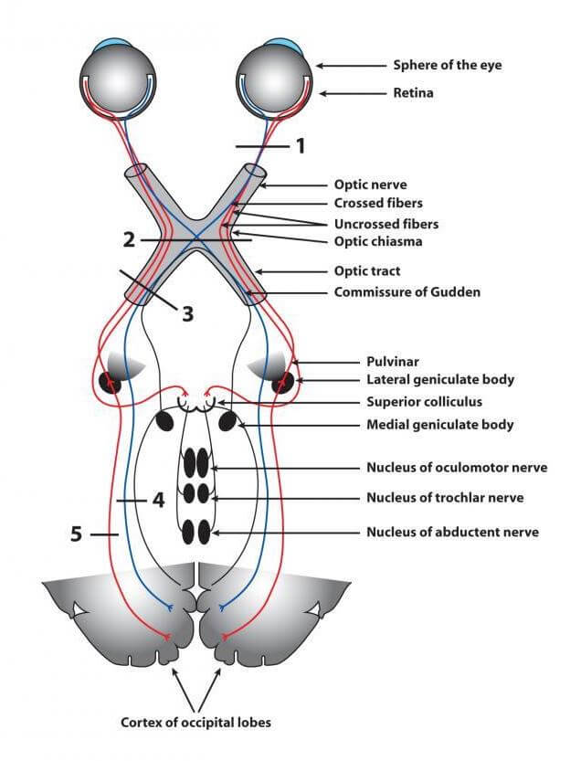
The optic nerve (CN II) is formed by the union of axons from the retinal ganglion cells and glial cells in the retina. The optic nerve passes through the orbit within the dural sheath and within the cone of muscles.
After formation, the optic nerve leaves the orbit via the optic canal, which is situated in the body of the sphenoid bone, to enter the middle cranial fossa. Here it lies superior to the ophthalmic artery.
Within the middle cranial fossa, the optic nerves from each eye run posteromedially to unite at the optic chiasm. The optic chiasm is located immediately below the hypothalamus and in close proximity to the pituitary gland. Fibres from the nasal (medial) half of the retina cross over at this point, forming the optic tracts.
The left optic tract, therefore, contains fibres from the left temporal retina and the right nasal retina. The right optic tract contains fibres from the right temporal retina and the left nasal retina. Each optic tract passes from the posterolateral angle of the chiasm, running lateral to the cerebral peduncle and medial to the uncus of the temporal lobe before reaching the lateral geniculate nucleus (LGN) in the thalamus.
The LGN acts as a relay centre and sends axons through the optic radiation to the primary visual cortex in the occipital lobe. The upper optic radiation carries fibres from the superior retinal quadrants (which corresponds to the lower half of the visual field) and travels through the parietal lobe. The lower optic radiation carries fibres from the inferior retinal quadrants (which corresponds to the upper half of the visual field) and travels through the temporal lobe.

The anatomy of the optic nerve and visual pathway
Function of the optic nerve
The optic nerve transmits all visual information including brightness perception, colour perception and visual acuity.
Examination of the optic nerve
The examination of the optic nerve involves the assessment of:
- Visual acuity
- Visual fields
- The retina
Visual acuity should be tested in each eye separately, and the patient’s vision should be corrected with eyeglasses or a pinhole before assessment. A Snellen chart is usually used, and the patient asked to read progressively smaller letters until their level of acuity is ascertained.
Visual fields are assessed by confrontation. The patient should cover one eye while the examiner tests the opposite eye. The examiner wiggles a finger in each of the four quadrants and asks the patient when it can be seen moving in the periphery. The examiner’s visual fields are used as a baseline for comparison and should, therefore, be normal.
Finally, the retina is assessed by fundoscopy.
Lesions of the optic nerve
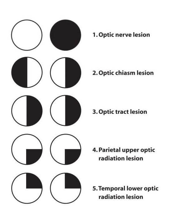
Lesions of the optic nerve (i.e. peripheral to the optic chiasm) cause ipsilateral monocular visual loss.
Causes of optic nerve lesions include:
- Optic neuritis, e.g. multiple sclerosis
- Optic nerve compression, e.g. ocular tumour
- Optic nerve toxicity, e.g. ethambutol or methanol toxicity
- Optic nerve trauma, e.g. orbital fracture
Lesions of the optic chiasm
The optic chiasm is located immediately below the hypothalamus and in close proximity to the pituitary gland. Enlargement of the pituitary gland can, therefore, affect the functioning of the optic nerve at this point.
At the optic chiasm fibres from the nasal (medial) half of the retina cross over to form the optic tracts. It is these fibres that are most affected by compression at the optic chiasm, and it produces a visual defect affecting peripheral vision in both eyes known as bitemporal hemianopia.
Causes of optic chiasm lesions include:
- Pituitary tumour (most common cause)
- Craniopharyngioma
- Meningioma
- Optic glioma
- Internal carotid artery aneurysm
Lesions of the optic tract
Homonymous hemianopias are caused by lesions of the optic tract.
Fibres from the nasal (medial) half of each retina cross over at the optic chiasm to form the optic tracts. The left optic tract, therefore, contains fibres from the left temporal retina and the right nasal retina. The right optic tract contains fibres from the right temporal retina and the left nasal retina.
A right-sided optic tract lesion will, therefore, cause loss of the left temporal and right nasal fields. A left-sided optic tract lesion will cause loss of the right temporal and left nasal fields.
Lesions of the optic radiation
Homonymous quadrantanopias are caused by lesions of the optic radiation.
Each optic tract passes from the posterolateral angle of the optic chiasm, running lateral to the cerebral peduncle and medial to the uncus of the temporal lobe before reaching the lateral geniculate nucleus (LGN) in the thalamus.
The LGN acts as a relay centre and sends axons through the optic radiation to the primary visual cortex in the occipital lobe. The upper optic radiation carries fibres from the superior retinal quadrants (which corresponds to the lower half of the visual field) and travels through the parietal lobe. The lower optic radiation carries fibres from the inferior retinal quadrants (which corresponds to the upper half of the visual field) and travels through the temporal lobe.
Therefore, temporal lobe lesions can cause superior homonymous quadrantanopias and parietal lobe lesions can cause inferior homonymous quadrantanopias.

Summary of the lesions of the optic nerve and visual pathway
Thank you to the joint editorial team of www.mrcemexamprep.net for this article.


